Home

listă Alinia Exemplu sarcomere structure microscope Incredibil laudă administrator

Cancer Histology Core

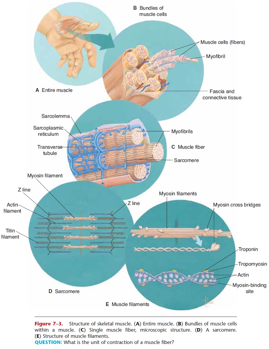
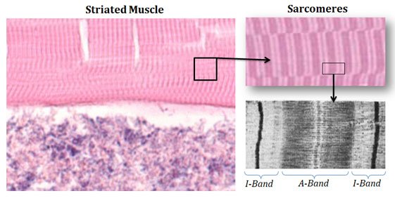
Muscle Fiber- Microscopic Structure

Solved Purpose: The purpose of this experiment is to observe | Chegg.com

Sarcomere - Definition, Structure, Function and Quiz | Biology Dictionary

Transmission Electron Micrograph- Sarcomere Structure (Partially Contracted  Muscle) Diagram | Quizlet
Transmission Electron Micrograph- Sarcomere Structure (Partially Contracted Muscle) Diagram | Quizlet

Polarization-resolved microscopy reveals a muscle myosin motor-independent  mechanism of molecular actin ordering during sarcomere maturation | PLOS  Biology
Polarization-resolved microscopy reveals a muscle myosin motor-independent mechanism of molecular actin ordering during sarcomere maturation | PLOS Biology

Muscle tissue - Knowledge @ AMBOSS

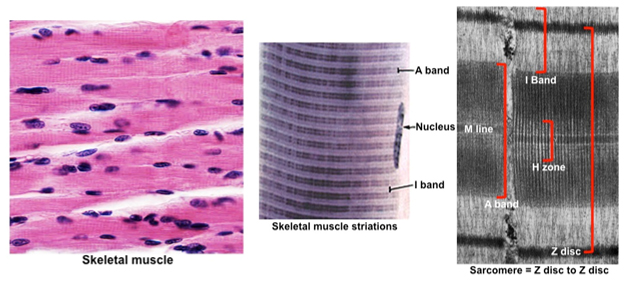
Muscle Lab
Muscle Lab

Electron microscopy image of a striated muscle sarcomere. Most of the... |  Download Scientific Diagram
Electron microscopy image of a striated muscle sarcomere. Most of the... | Download Scientific Diagram

The Titin/Telethonin Complex

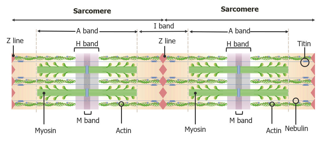
Sarcomere - Wikipedia

Titin stiffness modifies the force-generating region of muscle sarcomeres |  Scientific Reports
Titin stiffness modifies the force-generating region of muscle sarcomeres | Scientific Reports

The molecular basis for sarcomere organization in vertebrate skeletal  muscle - ScienceDirect
The molecular basis for sarcomere organization in vertebrate skeletal muscle - ScienceDirect

Sarcomere structure. A) Electron microscope image revealing the... |  Download Scientific Diagram
Sarcomere structure. A) Electron microscope image revealing the... | Download Scientific Diagram

Polarization-resolved microscopy reveals a muscle myosin motor-independent  mechanism of molecular actin ordering during sarcomere maturation | PLOS  Biology
Polarization-resolved microscopy reveals a muscle myosin motor-independent mechanism of molecular actin ordering during sarcomere maturation | PLOS Biology

Schematics of sarcomere and M-band structure derived from electron  microscopy of muscle [1],[2 ].
Schematics of sarcomere and M-band structure derived from electron microscopy of muscle [1],[2 ].

330 Electron Microscopy and the The Sliding Filament Model of Skeletal  Muscle Contraction - YouTube
330 Electron Microscopy and the The Sliding Filament Model of Skeletal Muscle Contraction - YouTube

Pradeep's Homepage
Pradeep's Homepage

a Electron micrograph of a skeletal muscle sarcomere, demonstrating... |  Download Scientific Diagram
a Electron micrograph of a skeletal muscle sarcomere, demonstrating... | Download Scientific Diagram

Figure 5 from The organization of titin filaments in the half-sarcomere  revealed by monoclonal antibodies in immunoelectron microscopy: a map of  ten nonrepetitive epitopes starting at the Z line extends close to
Figure 5 from The organization of titin filaments in the half-sarcomere revealed by monoclonal antibodies in immunoelectron microscopy: a map of ten nonrepetitive epitopes starting at the Z line extends close to

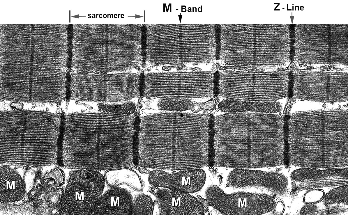
Skeletal Muscle Contraction | Concise Medical Knowledge

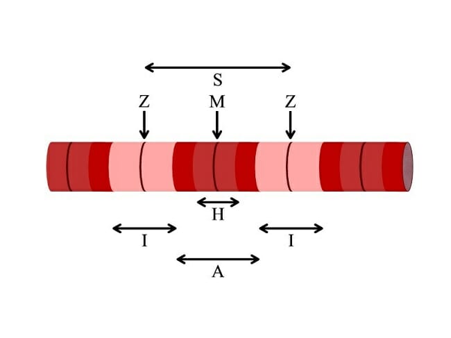
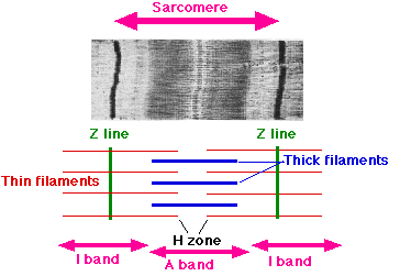
Difference between A-Band and I-Band - Sarcomere | EasyBiologyClass

Human Structure Virtual Microscopy

a) Electron micrograph showing a whole sarcomere from fish muscle in... |  Download Scientific Diagram
a) Electron micrograph showing a whole sarcomere from fish muscle in... | Download Scientific Diagram